 Cellular and Molecular Immunology
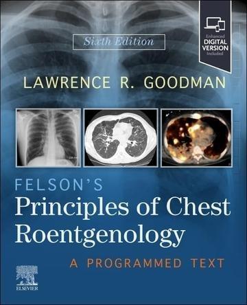
Felson's Principles of Chest Roentgenology
Wydawca: Elsevier
A programmed English-language tutorial in chest radiograph interpretation, combining anatomy, a systematic search pattern, differential diagnosis, ICU imaging, safety, quizzes, and over 550 images.
Gdzie kupić: ebook, audiobook
Epub, Mobi, PDF, MP3
This sixth edition uses programmed learning to teach the interpretation of chest radiographs and related thoracic imaging. It is intended for medical students, residents, radiologists, and clinicians who order or read chest x-rays. The approach breaks a complex visual task into a sequence of observations, questions, and answers, allowing readers to work at their own pace while repeatedly applying the same organized method.
The book begins with the formation of radiographic images, basic chest anatomy, and a systematic search pattern. Readers learn how to examine technical quality, compare structures, identify an abnormal sign, and decide which diagnostic possibilities deserve consideration. Emphasis remains on findings encountered in everyday practice, so the material connects foundational anatomy with practical recognition of disease.
A stepwise chapter organization encourages active participation. Rather than presenting long blocks of theory alone, the text asks the reader to interpret images, commit to an answer, and then compare that judgment with an explanation. Multiple-choice questions, quizzes, annotated radiographs, line drawings, cartoons, and clinical pearls reinforce key concepts and expose common errors. Humor and short comments are used to maintain attention without replacing the diagnostic reasoning.
The revised edition includes many new or upgraded images and adds a chapter devoted to imaging in the intensive care unit. It also provides new review questions and directs readers to the 2024 Fleischner Society glossary of terms for thoracic imaging. These updates place the established method in a current clinical context and support consistent use of terminology.
More than 550 radiographs form the visual core of the book. When appropriate, chest x-rays are correlated with CT, PET, or MR images so that a two-dimensional finding can be understood in relation to anatomy and other modalities. The comparisons help readers see why a sign appears as it does and how additional imaging may narrow the differential diagnosis.
The discussion also covers radiation safety and the principles needed to understand how technique affects image appearance. By combining image interpretation, anatomy, differential diagnosis, safety, and repeated self-assessment, the volume functions as a self-directed tutorial and a review resource. Its aim is to establish a reproducible reading habit that can be applied to routine films, urgent studies, and more complicated cases rather than to teach memorization of disconnected signs.
Dodatkowe informacje
Kategoria: Medycyna
Wydawca: Elsevier
Data pierwszego wydania: 1 stycznia 2027
Liczba stron: 300
Tytuł oryginalny: Felson's Principles of Chest Roentgenology
ISBN: 9780443287091
Język: en
Okładka: Miękka
Wydanie: 6
Reklama
Komentarze i recenzje
Zaloguj się, aby dodać komentarz lub ocenę.
Inne książki autora
Brak innych książek tego autora.
Inne książki w tej kategorii
Reklama
Brak ocen, komentarzy i recenzji.